Brain Imaging Technologies
The Alzheimer’s Hub of Hope has four sections: Heroes, Highlights, Headlines and Helpers/Caregivers. This post is aligned to the headlines section.
I’ve received requests to discuss the pro’s and con’s of the three recently FDA approved Alzheimer’s treatments: Donanemab, aducanumab (Aduhelm) and lecanemab (Leqembi). As I researched the topic it became clear to me that it is very complicated and I would need more time to do it justice. Not only is there controversy in the medical community regarding whether the drugs should be approved, there is possible regulator misconduct (e.g. fraud?) that spawned congressional hearings. I’ll post on it sometime in the future. Thanks to the subscribers who requested the topic and to subscriber Dave who sent me reference materials.
Some of you may have heard of Dr. Daniel Amen from his PBS specials, 12 New York Times best selling books or his affiliation with the NFL and their concussion protocols. He is known for the following quote that I pulled from the Amen Clinic website and slightly tweaked.
Unfortunately, psychiatry remains the only medical field that rarely looks at the organ it treats. But without functional brain imaging tools, clinicians will never be able to know the underlying brain patterns of the patients they treat, so they are handicapped to throw medicated-tipped darts in the dark at their patients.
Dr. Amen has a point. It seems self-evident that you’d want to see the physical body part that you are treating and how it functions. To that end, I’ve documented the brain scans/imaging terms I’ve seen in my research and how they are used in Alzheimer’s. Unfortunately, they are almost always used to help diagnose and confirm AD and very rarely are their results used to treat an AD sufferer. Not using this information may be a symptom of the profession’s "Diagnose and Adiós" approach I posted about here.
I was able to identify that Dr. Nate Bergman, one of my Heroes of Alzheimer’s, uses qEEG/brain mapping (see below) as part of his functional approach to treating dementia. Brain mapped data is combined with other assessment information such as blood work, and health history to create a unique and precise treatment plan for the sufferer.
Dr. Bergman isn’t the only person using scan data to help with treatment for AD but he’s one of the few I could find via 90 minutes of Google, DuckDuckGo and ChatGPT searches which tells me how unusual it is.
Below, I describe scanning and imaging technologies and bolded what each scan identifies in the brain to highlight the breadth and diversity of their functions. Scans can identify brain: atrophy, structural changes, abnormalities, activity, blood flow, blood oxygenation, glucose metabolism, neuronal dysfunction, amyloid plaques, blood vessel health, and electrical activity. This seems like a lot to me. I would hope in the future the profession would be more on-board using this information to develop treatment protocols.
Brain Scanning Technologies
qEEG (Quantitative Electroencephalography) / Brain Mapping
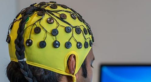
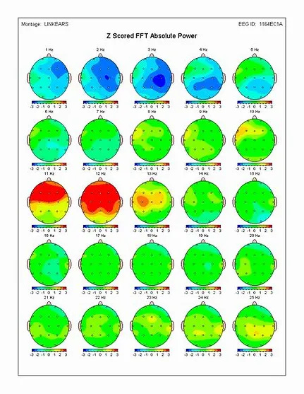
qEEG involves recording electrical activity from the scalp using electrodes, typically in the form of an electroencephalogram (EEG). The EEG measures the electrical signals produced by neurons in the brain. These electrical signals are brain waves such as delta, theta and alpha. The “q” in qEEG quantifies and analyzes the EEG data to provide information, a brain map, about the brain's electrical patterns and frequencies. It is often employed in clinical settings to study brain function, identify abnormalities, and assist in the diagnosis of various neurological and psychiatric conditions.
Neurofeedback has been used with qEEG to train patients to control their brain waves resulting in reduced anxiety, depression, PTSD and other symptoms. I’ve included reference materials at the bottom of the post for those who want more information on qEEG and brain mapping.
Below are images of the mechanics of qEEG and a brain map produced from this technology.
MRI (Magnetic Resonance Imaging): MRI is commonly used to detect structural changes in the brain. It can reveal atrophy (shrinkage) in specific regions, such as the hippocampus, which is often affected in the early stages of Alzheimer's.
CT Scan (Computed Tomography)
These are also referred to as CAT (Computerized Axial Tomography) scans.
CT scans can detect structural abnormalities and atrophy in the brain. However, MRI is generally preferred for Alzheimer's diagnosis due to its superior ability to visualize soft tissues.
Functional Magnetic Resonance Imaging (fMRI)
Is a neuroimaging technique that measures and maps brain activity by detecting changes in blood flow and oxygenation. It is a non-invasive method that provides insights into the functional organization of the brain by examining which areas are activated during various tasks or while the brain is at rest.
PET Scan (Positron Emission Tomography)
PET scans, particularly those using a radiotracer called FDG (fluorodeoxyglucose), can show patterns of glucose metabolism in the brain. In Alzheimer's patients, there is typically reduced glucose metabolism in certain brain regions, indicating areas of neuronal dysfunction. Another PET scan using a radiotracer specific to amyloid plaques (a hallmark of Alzheimer's) can visualize their accumulation in the brain.
Note: you may want to check out my Alzheimer's May Be Diabetes of the Brain post since it discusses how some AD sufferer brains don’t use glucose (sugar) effectively but can obtain their required energy from ketones. A PET scan can confirm which regions of the brain are negatively impaired by not metabolizing glucose and may benefit from ketones.
SPECT Scan (Single Photon Emission Computed Tomography)
SPECT scans can be used to assess blood flow in the brain, providing information about brain function. Like PET, SPECT can be employed to evaluate changes associated with Alzheimer's disease. SPECT allows physicians to look deep inside the brain to observe three things: areas of the brain that work well, areas of the brain that work too hard, and areas of the brain that do not work hard enough.
This is Dr. Amen’s go to brain imaging tool. Per the Amen Clinic’s website he treats brain injuries and other brain maladies but Alzheimer’s was not listed. You may want to check out Dr. Amen's NFL work using SPECT here.
Cerebral Angiography
This type of angiography focuses on the blood vessels in the brain. It is commonly used to evaluate conditions such as aneurysms, arteriovenous malformations, and other vascular abnormalities in the brain.
near infrared spectroscopy (NIRS)
Is a non-invasive optical technique that uses near-infrared light to measure changes in the absorption of light by biological tissues. This method is commonly employed to monitor oxygen levels and blood flow in various tissues, particularly in the brain and muscles. NIRS is widely used in medical and research applications to study tissue oxygenation, assess cerebral perfusion, and monitor changes in regional blood flow during tasks or interventions.
qEEG / Brain Mapping Reference Materials
qEEG Brain Mapping: What information does it give you?
How to Interpret Your qEEG Brain Map.
EEG Monitoring Approaches to Predict Learning and Memory Changes in Early Alzheimer’s Disease
Brain map qeeg (near the bottom of the page)


